601-608, Vedanta complex, Usmanpura, Ahmedabad – 13
601-608, Vedanta complex, Usmanpura, Ahmedabad – 13
The ACL is one of the major ligaments in the knee that connects the thigh bone (femur) to the shin bone (tibia). It is the main of the four ligaments stabilizing knee joint. When an injury occurs in the knees during sports, motorbike accident, two wheeler accident or in domestic twisting injuries – this is the ligament that is commonly torn. This injury is very common and its treatment is a popular topic of medical literature.
The ACL is important during daily activities like walking on uneven ground, on slippery surfaces, during brisk walking, dancing, running/jogging. It is absolutely critical for the stability of the knee during sports that require cutting and pivoting, such as tennis, badminton, cricket, soccer, football and basketball. What does the anatomy of the ACL look like?
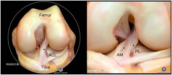
Although the ACL is referred to as one ligament, it consists of two functional bundles. These two bundles are named for the place where they attach on the leg bone (tibia). There is an Anteromedial (AM) bundle, which inserts more towards the front and towards the inside of the leg bone (tibia). The posterolateral (PL) bundle inserts most towards the back and towards the outside of the tibia.


1. Normal ACL
2. Ruptured ACL
3. Femoral end Avulsion
4. Stretched Dysfunctional ACL
The ACL provides stability to the knee, while also allowing for normal knee movement. The AM bundle is tight when the knee is bent and provides stability in the forward direction. The PL bundle is loose when the knee is bent, and allows for rotation of the knee. When the knee is straight the two bundles are parallel to each other, but when the knee is bent the two bundles cross each other. Although the two bundles have slightly different functions, the bundles do not work independently, but rather they work together to keep the knee stable while still allowing you to jump, run and play sports.
ACL tears are very common. The highest occurrence is in individuals between 15 to 25 years of age who participate in pivoting and cutting sports. However, ACL tears can occur at all ages and in all sporting activities after either contact or non-contact injuries. Usually an ACL tear occurs during sporting activities, two wheeler vehicular accident or may be in domestic accidents. They can be torn in a sudden pivoting or cutting motion, or planting of the foot while the rest of the body turns.

Patients frequently report “hearing a pop” at the time of injury and often have large swelling and pain soon after, in the injured knee.
Sometimes in day to day activities patient with torn ACL experience frequent “giving Way” (Subluxate) leading to periodic increase in the pain in the injured knee.
They feel less confident on their injured knee in brisk walking, walking on uneven surface, walking on wet or slippery surfaces.
They are uncomfortable in Jumping down from the small height like few steps and not willing to run/jog even for small distances.
Negotiating staircase down is often difficult for them. Some of them feel unexplained pain of exertion.
If they have been having meniscus tear with the ACL tear they can experience episodes of ”locking of knee” (not able to move knee joint) lasting from few minutes to few hours to few days. In short patients of ACL are otherwise able to lead near normal life with significant restrictions.

This is done through a thorough history of your injury as well as through a variety of physical exams which include anterior drawer test, Lachman’s, pivot shift test each of these tests aids in determining the functional status of the ACL. The physical examination in clinic is used to make the provisional diagnosis.
MRI scans are used to image the ACL, confirm the diagnosis and evaluate for other possible injuries, like meniscus tears.
I also take x-rays of the knee. You cannot see the ACL on x-ray, but I do this to make sure there is no problem with the bones, such as a fracture.
No, it does not heal. This ligament mostly being made of fibres like elastic tissue, both ends of the torn ligament move away from each other gradually and eventually there will be empty space between two torn ends which cannot bridge naturally leaving it unhealed.
There are some middle to old aged patients who are able to function without an intact ACL. These patients modify their lifestyle by eliminating aggressive normal activities that require pivoting and cutting. However, sometimes during everyday activities the ACL-deficient knee can buckle or “give way” (Subluxate) resulting in painful episodes with swelling. In short one has to restrict the life significantly so as to not provoke the episode of the giving way.
Importantly, there is a risk of damage to the menisci (the cartilage shock absorbers) and articular cartilage (the slippery gliding surface on the ends of the bones) with each subluxation event. This damage can lead to degenerative arthritis and subsequent meniscus tears. These tear of meniscus and damage to articular cartilage is largely irrecoverable even when patient opt for surgery at the later date. Because of these concerns a majority of active and young patients elect to undergo ACL surgery when the ligament tears.
In general, there are fewcriteria that must be met before the ACL can be surgically reconstructed:
1, Swelling in the knee must go down to near-normal levels
2, Range-of-motion (bending and straightening) of the injured knee must be nearly 50% of the uninjured knee.
3, Good Quadriceps muscle strength must be present. This means that while lying flat on your back you should be able to raise your leg off the ground while holding it is straight. I call this a “straight leg raise”.
Usually it takes a few weeks after injury before ACL reconstruction can be performed.
The mode of injury like domestic accident or vehicular accident. The velocity of the injury low, moderate or high velocity also play big role in the decision.
The presence of any associated injuries to the knee joint involving cartilage, Contusion of the marrow, meniscus, or other ligaments may change the time-frame for surgery.
It is very tailor made decision, considering various divers factors mentioned above, arrive at by your surgeon in your best interest, regarding the time of the surgery.
yes, recently trend and new technique of ACL repair is being performed in the very few selected suitable cases. ACL repair is typically being performed in relatively fresh ACL injuries of femoral end avulsion cases in young individuals. It is more suitable for cases of partial tear in the form of either AM or PL bundle only, with intact remaining bundle, but it can be performed in complete ACL femoral end avulsion also. However final decision of repair versus reconstruction will be taken by your doctor only during the surgery.

The goal of anatomic ACL reconstruction is to reproduce 60 – 80% of the native ACL attachment site area which is more than enough for all your activities including sporting activities.
The goal of reconstructing the ACL is to restore the native anatomy and function of the ACL. Reproducing anatomy is the primary principle in Orthopaedic, like in the treatment of broken bones. Therefore, since 2011, I have changed our approach to doing anatomic ACL reconstruction. I try to preserve the bone and soft tissue, which allows me to restore both ACL anatomy and function. I believe this will help protect the long-term knee health.
The Purpose of anatomic ACL reconstruction is to regain stability and return to pre-injury activity level& maintain long term knee health.
The surgical procedure itself takes between 60 and 90 minutes. You will be in the operating room between 90 and 120 minutes because I repeat the physical examination of your knee when you are in the operating room.
To reconstruct the ACL, you are given general anesthesia and arthroscopy is performed. This means I look inside the joint with a small camera using small punctures and very small delicate instrumentation.


– lateral portal which is puncture towards the outside of the knee is used to insert the arthroscope in the joint.
-medial portal which ispuncture towards the inside of the knee is used to insert highly specialised small instrument sin the joint for the procedure.
One inch long incision over the tibia is used to mostly harvest the graft and create the tibial tunnel through which graft is introduced to the joint and attach the new ACL to the tibia.
Occasionally, other incision in the upper part of the knee and near ankle is used to harvest the more graft if required. An additional incision is made on the outer aspect of the knee joint over the femur to help attach the new ACL to the femur.
When the camera (arthroscopy) is place inside the knee, I look carefully at the injured ACL. I determine where and how it is torn. It can be partially or completely torn, and torn from the top, in the middle or on the bottom. Sometimes, the ACL is still attached to the bones, but often it is stretched out and has lost its function.
The above picture on the left shows a normal ACL as seen during arthroscopy. In the second picture, the whole ACL is torn of the femur completely. In the 3rdpicture ACL has avulsed from the femoral end. In the 4th picture ACL is stretched and rendered dysfunction but structurally appears intact.


After the injured part and unusable part of ACL is carefully removed, the attachments (also named insertion sites) of the ACL on the bone can be seen clearly.
The insertion sites of the ACL is then carefully measured with a small ruler. These measurements will determine the size of the new ACL
A new ACL is created using “a graft”, which is tissue as mentioned earlier from your own body. The sizes of these grafts are based on your own body size body type and ACL size.


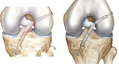
To attach the ACL graft to the bone with very precise and special instruments tunnels are drilled in the bone according to the size and length of the graft and then ACL graft is placed into the tunnels and fixed to the femur and tibia bones with a combination of special buttons, screws and sometimes with staples depending upon the need condition of the bone and graft.
These fixation options are discussed with you explaining the pros and cons of each fixation option before the surgery to help you to choose appropriate fixation mode On the X-ray of the knee we can see the tunnel through the femur and tibia.

The graft tissue comes from your own body (autograft)
Autograft options include different tendons from different muscles: Hamstrings Tendons, Quadriceps Tendon, and Patellar Tendon (BTB), peroneus longus tendon, hamstring tendon from opposite knee.
Advantages to autograft include no risk of disease transmission and potentially quicker healing of the new ACL.
Allograft (Tissues harvested from Dead people) options also include a variety of different tendons from different muscles: Hamstrings Tendons, Tibialis Anterior Tendon, Posterior Tibial Tendon, Patellar Tendon, Quadriceps Tendon, Achilles Tendon, and the Tensor Fascia Lata. But it is not available in india and illegal also. It also has inherent risk of disease transmission like hepatitis and HIV as well as prolonged healing time of ACL.
Typically the graft heals to the bone through bleeding created by drilling the tunnels. The type of fixation material also helps newly reconstructed ACL to heal in the tunnels. It will get its normal synovial covering in 3 months time then the blood supply from it will help in further naturalization process.
I carefully follow our patients after surgery and measure knee stability, as well as knee range of motion. In the picture below you see straightening of the legs and straight leg raise, bending of the knees, kneeling and strength of the quadriceps muscle.
After anatomic ACL reconstruction, most patients achieve excellent range-of-motion, typically equal to the other knee. These results are usually seen as early as 1 to 3 months after surgery.
Lorem ipsum dolor sit amet, consectetur adipiscing elit. Ut elit tellus, luctus nec ullamcorper mattis, pulvinar dapibus leo.
After anatomic ACL reconstruction, rehabilitation guidelines are usually as follows:
In general, these guidelines should be followed after ACL reconstruction. However, it is very important to realize that these guidelines may change depending on each patient. Your doctors may tailor a specific, individualized rehabilitation program depending on the number of surgeries you have had, accompanying ligament and meniscal injuries, your individual progress, and other factors that may impact the healing of you graft.
All guidelines should be followed closely because the new ACL needs time to heal. Remember, returning to sports before the graft is healed increases the chances of re-injury. Although you may feel “fine” earlier, the ACL graft takes about 9 months to heal.
“Why does an anatomical graft take 9 months to heal?” An anatomically placed ACL experiences same forces as normal ACL therefore, may be easier to re-rupture! Before it is fully ligaments (Can behave as native ACL) within the joint. This process is slow and cannot be hastened.
The anatomically placed ACL better restores normal knee function and it needs 9 months of time to become strong enough to withstand sporting stresses to enable you resume the sports activities.